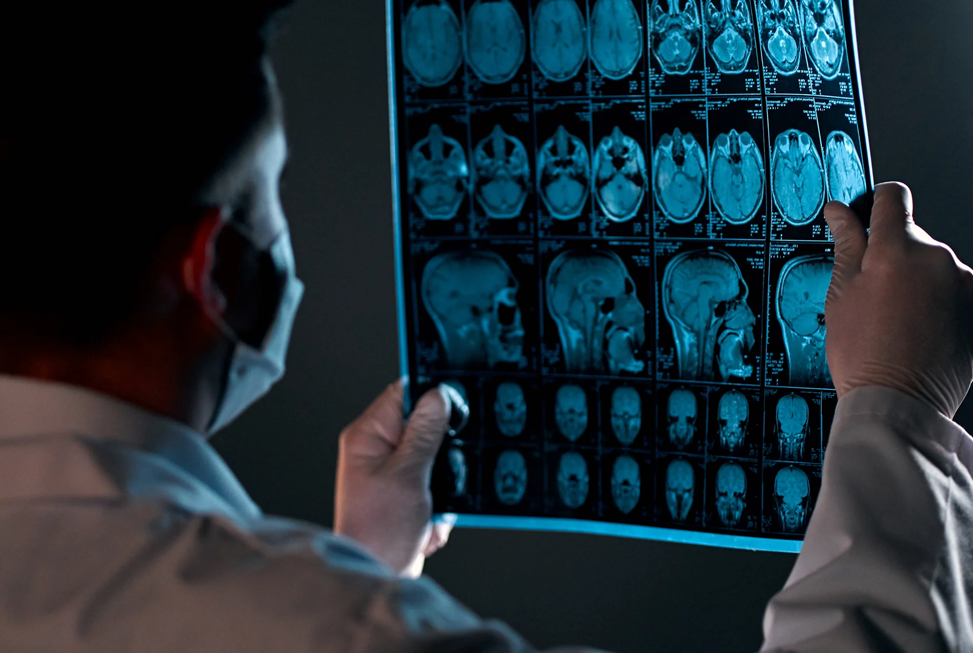
Patients often come to consultation focused on the change they want to see. They may be interested in a straighter or smaller nose or a more refined nasal bridge. What they sometimes don’t expect is how much of the planning happens before the surgery ever begins. Preoperative imaging in nasal surgery and septorhinoplasty in particular is one of the most valuable tools in that process. At Conderman Facial Plastic Surgery in Orinda, CA, it plays a role in surgical decision making for nearly every patient.
What Preoperative Imaging Actually Is
Preoperative imaging refers to the photos, measurements, and imaging studies captured before surgery to document your starting point and helps to guide the surgical plan. In facial plastic surgery, that typically includes:
- Standardized clinical photography – front, profile, oblique, base and top down views, taken with consistent lighting, distance, and head position
- Close-up detail views – focused on the specific features in question, such as the nasal tip, alar base
- Dynamic views – images captured in animation (smiling and while taking a breath in through the nose) to show how features move, not just where they are at rest
- CT imaging – when airway or bony structure matters, as in functional rhinoplasty or revision cases
- Three-dimensional surface imaging – where available, useful for evaluating volume, asymmetry, and contour in ways a flat photo cannot. This is especially useful for three dimensional anatomy of the nasal bones and bony vault as this has implications for osteotomy design (i.e. how the nasal bones will be addressed during nasal surgery)
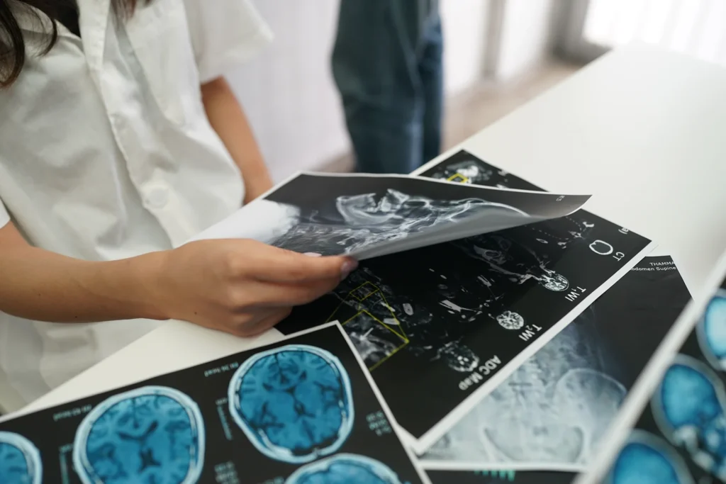
The point is not documentation for its own sake. Imaging plays a critical role in helping to design the surgical plan, aka the roadmap you and your surgeon use to agree on where you’re going/the goals for your procedure.
Why It Matters: Six Practical Reasons
- It reveals what a mirror can’t. Patients see themselves in the mirror daily, usually front-on and in motion. Standardized imaging shows angles you may not routinely evaluate yourself – the profile, the base of the nose, the top-down view. Asymmetries and structural issues that aren’t typically accounted for in daily life become obvious when documented consistently. In a previous case here, a patient’s mid-vault asymmetry and infra-tip imbalance were clearest on the top-down view – an angle she had never actually seen.
- It makes the conversation concrete. “It’s too big” or “it droops” are starting points, not surgical plans. Imaging lets you point at a specific feature, at a specific angle, and talk about it in a shared language. That reduces the gap between what you mean and what your surgeon understands.
- It sets realistic expectations. This may be the most important function. Imaging – and, where appropriate, digital simulation – helps show what is achievable given your anatomy and what is not. A nose can be refined; it cannot be replaced with someone else’s.
- It guides technique, not just aesthetics. For functional cases, imaging contributes directly to surgical decision making. A patient with nasal obstruction and visible asymmetry may need structural support – a spreader graft, septum repositioning, mid-vault reinforcement – rather than simple reduction. Knowing what the nasal anatomy is going to be preoperatively can change the surgical paln.
- It is a shared record of consent and care. Consistent, permission-based photography is part of a responsible plastic surgery practice. It documents the starting point, supports clinical decision-making, and – with your consent – allows your surgeon to share outcomes that help future patients understand what a procedure can realistically do.
Common Misconceptions
“Imaging is just before-and-after photos.”
Before-and-after photos are the output. The preoperative portion – standardized capture, measurement, and comparison – is the diagnostic work that makes those images meaningful.
“Simulation means I’ll get that exact result.”
Simulation is a communication tool, not a guarantee. It helps align expectations on direction and proportion. Final results depend on anatomy, healing, and technique.
“If I don’t like my photos, that’s a bad sign.”
Standardized photos are deliberately unflattering. Flat lighting, no expression, and direct angles are designed to reveal structure, not to flatter. They are clinical documents, not portraits.
“More imaging means more radiation or more cost.”
Most preoperative imaging in facial plastic surgery is photography. CT or Cone Beam imaging can also be used to assess the nasal anatomy and to guide the surgical plan
What to Expect at Your Consultation
- Standardized photography at the outset, in a consistent setup
- The photos and renderings will be reviewed together on screen, with the features you want addressed specifically discussed
- Discussion of which views matter for your particular goal, and why
- Simulation and renderings where appropriate, framed as a conversation about direction and proportion
- A written plan that references your documented starting point
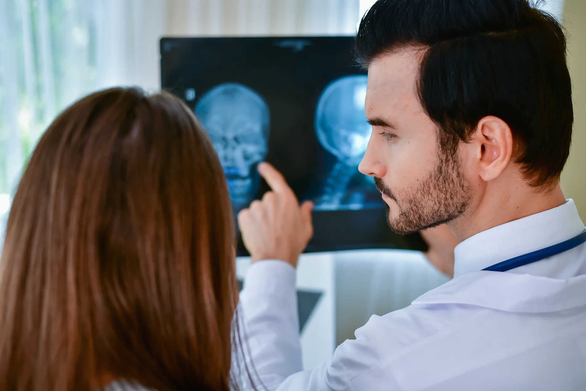
Bring reference photos if you have them – images of results you admire are useful, as long as the conversation stays anchored to what your own anatomy can support.
How Imaging Fits Into a Rhinoplasty Plan
- Document your baseline with standardized views
- Identify the structural and aesthetic elements driving your concerns
- Decide on technique based on anatomy, function and aesthetics
- Align on realistic goals through simulation and discussion
- Compare at each follow-up to track healing honestly
- Refine the plan at every stage based on what the images show
Frequently Asked Questions
Are the photos required?
Yes, for any surgical planning. Without a documented starting point, both the surgical plan and the follow-up assessment lose their reference.
What happens to my images?
They stay in your clinical record. Images are shared publicly only with your explicit permission, and identifying details are handled according to practice policy and privacy standards.
Can I see my photos?
Yes. Reviewing them together is part of consultation – that is often when patients first see their asymmetry clearly.
Is simulation accurate?
It is directional, not predictive. Use it to confirm that you and your surgeon are imagining the same change, not as a promise of a specific result.
Do I need a CT scan?
Dr. Conderman uses pre-operative imaging to help guide surgical decision making and this is an important part of the pre-operative process. This is especially important for significant airway obstruction, prior trauma, or revision surgery where bony detail matters.
The Bottom Line
Preoperative imaging is diagnostic tool and facilitates communication between patient and surgeon. Thorough pre-operative assessment of imaging turns vague concerns into specific plans, guards against unrealistic expectations, and lets both patient and surgeon track healing honestly over months.
If you are considering Rhinoplasty in Orinda, CA or the surrounding Bay Area, a thorough consultation – starting with proper imaging – is the first meaningful step.